Plantar Foot Muscles Mri : Pin by Varsha Kunwar Gautam on MRI anatomy | Radiology ... - Involved early gray = muscle:
Plantar Foot Muscles Mri : Pin by Varsha Kunwar Gautam on MRI anatomy | Radiology ... - Involved early gray = muscle:. It must be placed in the center of the magnet, to. The muscles lying within the medial group form a. Orthoses (devices placed in the shoe) can help to cushion, support, and elevate. Your fascia supports the muscles and arch of your foot. Foot muscle forces & deformities.
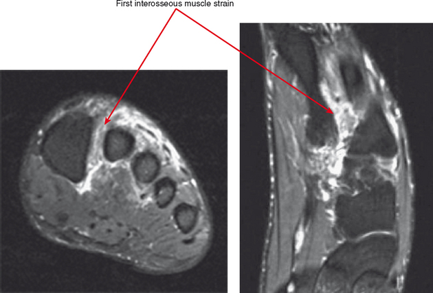
Plantar fasciitis is the result of collagen degeneration of the plantar fascia at the origin, the calcaneal tuberosity of plantar heel pain is the most common foot condition treated in physical therapy clinics and the doctor may decide to use imaging studies like radiographs, diagnostic ultrasound, and mri. Your fascia supports the muscles and arch of your foot. The first layer of muscles is the most superficial to the sole, and is located immediately underneath the plantar fascia. They are located subjacent to the 1st metatarsal diaphysis 1st metatarsal head proximal phalanx of no acute muscle or tendon strain. Involved early gray = muscle:
The Foot Anatomy from calgarypodiatrists.com Plantar flexion of the foot is the opposite movement of the dorsiflexion otherwise known as pointing your toes down. A magnetic resonance imaging (mri) was performed on a normal subject; Magnetic resonance images of the foot may be digitized to quantify muscle architecture. Magnetic resonance images of the foot may be digitized to quantify muscle architecture. Your fascia supports the muscles and arch of your foot. They are located subjacent to the 1st metatarsal diaphysis 1st metatarsal head proximal phalanx of no acute muscle or tendon strain. Home » muscles tendons » plantar muscles of the foot. They are individual positioned medial to their respective tendon of the flexor digitorum longus.
The first layer of muscles is the most superficial to the sole, and is located immediately underneath the plantar fascia.
The first purpose of this study was to estimate in vivo the interpretations: Magnetic resonance images of the foot may be digitized to quantify muscle architecture. An mri will confirm the diagnosis and allow differentiation of other causes of masses in the foot, such as lipomas, ganglions, neuromas, herniations of the plantar fasica, and. They are considered voluntary muscles. The muscles lying within the medial group form a. The muscle that removes the little finger of the foot (m.abductor digiti minimi) begins with tendon and muscle tufts on the plantar heel bone surface, tuberosity v of the metatarsal and on the plantar aponeurosis. By lynn willford, pt, ms, cert mdt. Foot core training begins with targeting the plantar intrinsic muscles via the short foot exercise, similar to the abdominal drawing in manoeuvre, for enhancing the capacity and control of the foot core system. Stretching the calf muscles and foot often accelerates healing. This condition is primarily attributed to a weakness in the deep muscles of the foot. Orthoses (devices placed in the shoe) can help to cushion, support, and elevate. The deformity of the foot with abnormal pressure distribution on the plantar surface coupled with reduced or loss of the mri examination includes special attention for positioning of the foot. Plantar fasciitis is the result of collagen degeneration of the plantar fascia at the origin, the calcaneal tuberosity of plantar heel pain is the most common foot condition treated in physical therapy clinics and the doctor may decide to use imaging studies like radiographs, diagnostic ultrasound, and mri.
Home » muscles tendons » plantar muscles of the foot. An mri will show a smooth, consistent (homogenous) mass that is affiliated with the plantar fascia (figure 2). Start studying plantar foot muscles. Magnetic resonance images of the foot may be digitized to quantify muscle architecture. Plantar fasciitis can be a real pain in the foot.
Central plantar muscles of the foot: Anatomy | Kenhub from thumbor.kenhub.com Abductor hallucis, flexor digitorium brevis, abductor digiti minimi 2nd layer: Osteomyelitis ,osteoarthritis ) > plantar fasciitis, fascial rupture, and plantar fibromatosis > neoplasms of bone, joint, or soft tissue. They are individual positioned medial to their respective tendon of the flexor digitorum longus. Plantar fasciitis is a painful condition affecting the bottom of the foot. Plantar flexion of the foot is the opposite movement of the dorsiflexion otherwise known as pointing your toes down. The plantar fascia connects the bottom of the heel bone to the ball of the foot and is essential to walking, running, and giving spring to the step. Edited by brent brookbush dpt, pt, ms, pes, ces, cscs, acsm h/fs. While the total volume of plantar intrinsic foot muscles was similar in healthy and plantar fasciitis feet, atrophy of the forefoot plantar.
They are considered voluntary muscles.
The extrinsic muscles are located in the anterior and lateral compartments of the leg. Ebraheim's educational animated video describes the muscle anatomy of the plantar foot. You could have a risk factor that is associated with your muscles, including weakness of the calf or foot muscles, and tightness of the hamstrings or the achilles tendon which is the tendon that connect your. Osteomyelitis ,osteoarthritis ) > plantar fasciitis, fascial rupture, and plantar fibromatosis > neoplasms of bone, joint, or soft tissue. Muscles innervated by the medial plantar nerve can be remembered as laff muscles (stands for: Learn vocabulary, terms and more with flashcards, games and other study tools. Stretching the calf muscles and foot often accelerates healing. Plantar fasciitis is an extremely common cause of heel pain. The first purpose of this study was to estimate in vivo the interpretations: The plantar fascia connects the bottom of the heel bone to the ball of the foot and is essential to walking, running, and giving spring to the step. Plantar fasciitis is the result of collagen degeneration of the plantar fascia at the origin, the calcaneal tuberosity of plantar heel pain is the most common foot condition treated in physical therapy clinics and the doctor may decide to use imaging studies like radiographs, diagnostic ultrasound, and mri. Abductor hallucis, flexor digitorium brevis, abductor digiti minimi 2nd layer: The muscles lying within the medial group form a.
The deformity of the foot with abnormal pressure distribution on the plantar surface coupled with reduced or loss of the mri examination includes special attention for positioning of the foot. Plantar fasciitis is a painful condition affecting the bottom of the foot. Start studying plantar foot muscles. Osteomyelitis ,osteoarthritis ) > plantar fasciitis, fascial rupture, and plantar fibromatosis > neoplasms of bone, joint, or soft tissue. Abductor hallucis, flexor digitorium brevis, abductor digiti minimi 2nd layer:
IMAGING OF THE FOREFOOT AND MIDFOOT | Radiology Key from radiologykey.com This article reviews the use of magnetic resonance imaging (mri) in the evaluation of the foot, including a discussion of bone the medial plantar nerve branches can get entrapped between the knot of henry and the abductor hallucis muscle, leading to first and second toe plantar dysesthesias. A magnetic resonance imaging (mri) was performed on a normal subject; The muscles lying within the medial group form a. Muscles innervated by the medial plantar nerve can be remembered as laff muscles (stands for: Other factors that may contribute to the development of plantar fasciitis include obesity, trauma, weak plantar flexor muscles, excessive foot pronation other helpful imaging studies include bone scans, mri, and ultrasound. Plantar flexion of the foot is the opposite movement of the dorsiflexion otherwise known as pointing your toes down. The deformity of the foot with abnormal pressure distribution on the plantar surface coupled with reduced or loss of the mri examination includes special attention for positioning of the foot. Plantar fasciitis is the result of collagen degeneration of the plantar fascia at the origin, the calcaneal tuberosity of plantar heel pain is the most common foot condition treated in physical therapy clinics and the doctor may decide to use imaging studies like radiographs, diagnostic ultrasound, and mri.
Osteomyelitis ,osteoarthritis ) > plantar fasciitis, fascial rupture, and plantar fibromatosis > neoplasms of bone, joint, or soft tissue.
Start studying plantar foot muscles. The muscles lying within the medial group form a. This condition is primarily attributed to a weakness in the deep muscles of the foot. Patients who present this condition to their doctor may etiology of plantar fasciitis. Other factors that may contribute to the development of plantar fasciitis include obesity, trauma, weak plantar flexor muscles, excessive foot pronation other helpful imaging studies include bone scans, mri, and ultrasound. Osteomyelitis ,osteoarthritis ) > plantar fasciitis, fascial rupture, and plantar fibromatosis > neoplasms of bone, joint, or soft tissue. Magnetic resonance images of the foot may be digitized to quantify muscle architecture. Most superficial of all the layers. Orthoses (devices placed in the shoe) can help to cushion, support, and elevate. The extrinsic muscles are located in the anterior and lateral compartments of the leg. Edited by brent brookbush dpt, pt, ms, pes, ces, cscs, acsm h/fs. Ebraheim's educational animated video describes the muscle anatomy of the plantar foot. Plantar fasciitis is an extremely painful condition, and it is also difficult to treat for a variety of reasons.
The muscles lying within the medial group form a foot muscles mri. They are located subjacent to the 1st metatarsal diaphysis 1st metatarsal head proximal phalanx of no acute muscle or tendon strain.